Patellar Luxation in Dogs
Learn about patellar luxation in dogs, including its symptoms, four grades, treatment options, and when surgery may be recommended.
Dr. Sofia Raldiris
DVM
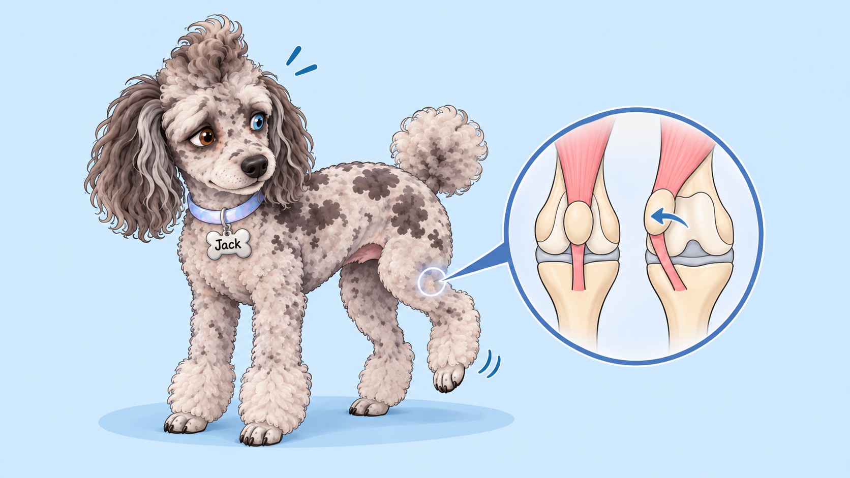
PATELLAR LUXATION IN DOGS
WHAT IS PATELLAR LUXATION?
Patellar luxation occurs when the kneecap (patella) slips out of its normal position in the knee joint. Normally, the patella glides along a groove at the end of the femur (thighbone), called the trochlear groove, which keeps it aligned as the knee bends and straightens.
Patellar luxation is classified by the direction the kneecap moves:
- Medial patellar luxation (MPL): The kneecap slips toward the inside of the leg. This is the most common form, especially in small and toy breeds.
- Lateral patellar luxation (LPL): The kneecap slips toward the outside of the leg. This is less common and occurs more frequently in larger dogs.
Most cases are developmental, resulting from abnormal bone alignment or soft tissue structure. Contributing factors include a shallow trochlear groove, abnormal femoral or tibial alignment, and improper attachment of the patellar tendon. Although trauma can cause luxation, it often develops early in life.
🔍 SIGNS OF A PATELLAR LUXATION
The most recognizable sign of medial patellar luxation (MPL) is an intermittent skipping gait.
Your dog may suddenly lift a hindlimb for several steps and then return to normal. When the kneecap slips out of its groove, your dog may have difficulty fully extending the knee. Once it returns to position, walking often returns to normal.
Other signs may include:
- Intermittent or persistent hindlimb lameness.
- Difficulty jumping, climbing stairs, running, or playing.
- Abnormal or bowlegged stance in advanced cases.
- Reduced knee extension.
- Muscle loss or weakness in the affected hindlimb.
Some dogs show no obvious symptoms, and luxation is discovered during a routine veterinary examination.
Even without obvious pain or limping, repeated luxation can cause cartilage damage, inflammation, osteoarthritis, and progressive limb deformity.
Sudden or severe lameness warrants prompt veterinary evaluation, as other conditions, including cranial cruciate ligament injuries, may also be responsible.
🌐 Learn more about CCL injuries in dogs in our article on Ruptured Cranial Cruciate Ligaments (CCL) in Dogs.
🩺 DIAGNOSIS
Patellar luxation is diagnosed through a physical and orthopedic examination. Your veterinarian assesses kneecap stability, determines the grade, and checks for other causes of lameness.
Radiographs (X-rays) may be recommended to evaluate joint and bone alignment. Advanced imaging, such as CT, may be needed in more complex cases to assess skeletal deformities and plan surgery.
Both hindlimbs should be evaluated, as the condition commonly affects both knees, even when symptoms appear on only one side.
Patellar Luxation Grades
Patellar luxation is classified into four grades based on how easily the kneecap dislocates, whether it returns to its normal position, and the degree of joint instability.
The grade helps guide treatment decisions, but your dog's symptoms, age, activity level, limb alignment, and overall orthopedic health must also be considered.
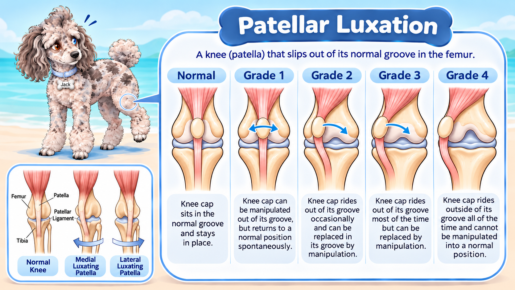
Grade I: Mild
The kneecap can be manually displaced but immediately returns to its normal position when released.
Dogs typically have no symptoms or only occasional signs.
Treatment: Surgery is usually unnecessary. Maintaining a healthy weight, controlled exercise, joint supplements, and regular veterinary monitoring are generally sufficient.
Grade II: Moderate
The kneecap slips out of its groove more easily, sometimes during normal movement. It may return on its own or require manual repositioning.
Dogs may experience intermittent skipping, occasional lameness, or reduced use of the affected leg.
Treatment: Surgery is not always necessary. Mild cases can often be managed with weight control, controlled exercise, joint supplements, and regular monitoring. Recurrent lameness or worsening instability may warrant surgery.
Grade III: Severe
The kneecap remains dislocated most of the time but can be manually repositioned. However, it quickly slips out again.
Dogs may develop persistent lameness, abnormal limb alignment, and difficulty fully extending the knee.
Treatment: Surgery is generally recommended to restore stability, improve limb function, and help prevent further joint damage.
Grade IV: Very Severe
The kneecap is permanently dislocated and cannot be manually repositioned.
Dogs may develop significant limb deformity, a crouched gait, and an inability to fully extend the knee. Longstanding cases may involve severe femoral and tibial deformities, making correction more complex.
Treatment: Surgery is generally recommended when feasible. Advanced cases may require more extensive bone-correcting procedures, and outcomes depend on the severity and duration of the condition.
🦴 TREATMENT
Conservative management
Conservative management may be appropriate for dogs with mild luxation, minimal symptoms, or those that are not good surgical candidates.
Management may include:
- Weight Management: Maintaining a healthy weight reduces joint stress.
- Controlled Exercise: Low-impact activity helps maintain muscle strength without excessive strain.
- Physical Rehabilitation: Targeted exercises improve strength, balance, and mobility.
- Medication: Veterinarian-prescribed anti-inflammatory or pain medication can help manage discomfort and osteoarthritis.
- Joint Supplements: May support joint health as part of an individualized care plan, although benefits vary.
These measures help manage symptoms but do not correct the underlying anatomical abnormalities. Dogs with significant instability or worsening lameness should be reassessed for possible surgical correction.
🛒Looking for products to support your pet’s joint health and mobility? Explore some of our recommended joint health products below.
When Is Surgery Recommended?
Not every dog with patellar luxation needs surgery. The decision depends on the grade of luxation, severity of symptoms, and impact on mobility. Surgery is generally recommended for Grade III and IV cases and may be considered for Grade II dogs with recurrent lameness, pain, or worsening instability.
Other reasons to consider surgery include persistent limping, progressive limb deformity, and difficulty with normal activities.
Surgery aims to stabilize the kneecap and restore proper alignment. Depending on the underlying abnormalities, procedures may include:
- Tibial Tuberosity Transposition: Repositions the patellar tendon attachment to improve kneecap alignment.
- Trochleoplasty: Deepens the femoral groove to help keep the kneecap in place.
- Soft Tissue Balancing: Releases or tightens surrounding tissues to improve stability.
- Corrective Osteotomy: Realigns the femur or tibia in cases with significant bone deformities.
Early evaluation and treatment can help prevent progressive joint damage and limb deformity. Even after successful surgery, some dogs may need ongoing arthritis management to maintain comfort and mobility.
What Happens If Left Untreated?
Mild cases may remain stable for years, but more severe or symptomatic cases can lead to progressive joint damage and reduced mobility.
Potential complications include:
- Progressive Joint Damage: Repeated kneecap displacement causes abnormal joint movement, cartilage damage, and inflammation, potentially leading to osteoarthritis, chronic pain, and reduced mobility.
- Abnormal Limb Development: In growing dogs, persistent luxation can cause abnormal femoral and tibial alignment, leading to progressive limb deformity and more complicated surgical correction.
- Muscle Weakness and Reduced Mobility: Reduced use of the affected leg can cause muscle loss and weakness. Abnormal gait may also place additional stress on other joints and muscles.
- Increased Risk of Other Knee Injuries: Altered knee mechanics may contribute to additional orthopedic problems, including cranial cruciate ligament disease.
Sudden or worsening lameness warrants veterinary evaluation to rule out additional injuries rather than assuming patellar luxation is the only problem.
Osteoarthritis
Osteoarthritis is common in dogs with patellar luxation, regardless of treatment. Joint instability and inflammation can cause progressive cartilage damage over time.
Long-term management focuses on maintaining comfort and mobility through healthy weight management, low-impact exercise, physical rehabilitation, joint supplements, and veterinarian-prescribed pain medication.
🌐 Learn more about long-term joint care in our article on Arthritis in Dogs and Cats.
🛒 RECOMMENDED JOINT HEALTH PRODUCTS
Joint-health products may help support your dog’s mobility and overall joint health. We’ve selected some recommended options below.
🌐 View Our Recommended Joint Health Products
Remember: These products do not correct patellar luxation or replace veterinary treatment. Your veterinarian can help determine the most appropriate management plan for your dog.
The Bottom Line
Not every dog with patellar luxation needs surgery. However, frequent lameness, worsening symptoms, and moderate to severe instability are important reasons to consider surgical correction.
If your dog regularly skips while walking, holds up a hind leg, or struggles with normal activities, schedule an orthopedic evaluation. Identifying the grade and assessing joint health can help determine the best treatment to protect your dog's long-term comfort and mobility.
❓HAVE MORE QUESTIONS?
We’re here to help you keep your furbaby happy, healthy, and thriving! ❤️
For additional pet health information, visit Veterinary Partner by VIN—a trusted resource with veterinarian-written articles.
🔎 Tip: Search “VIN + your question” on Google for easier results, as the website’s search feature can be a little tricky.
📞 And remember, calling us is always free!
If you have questions or concerns about your pet, please don’t hesitate to reach out. We’re happy to help. 🐾